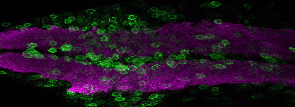
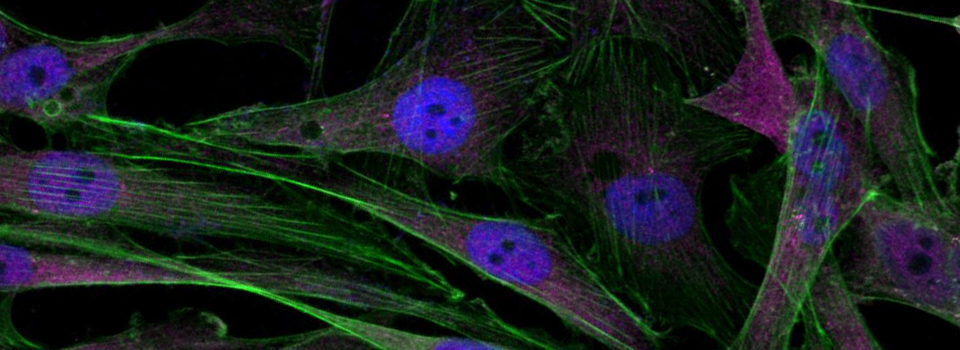
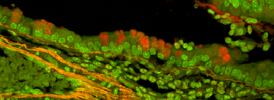
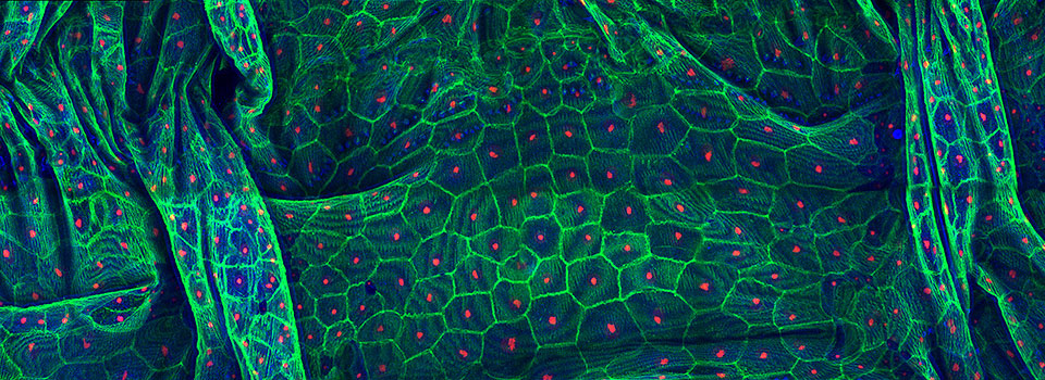

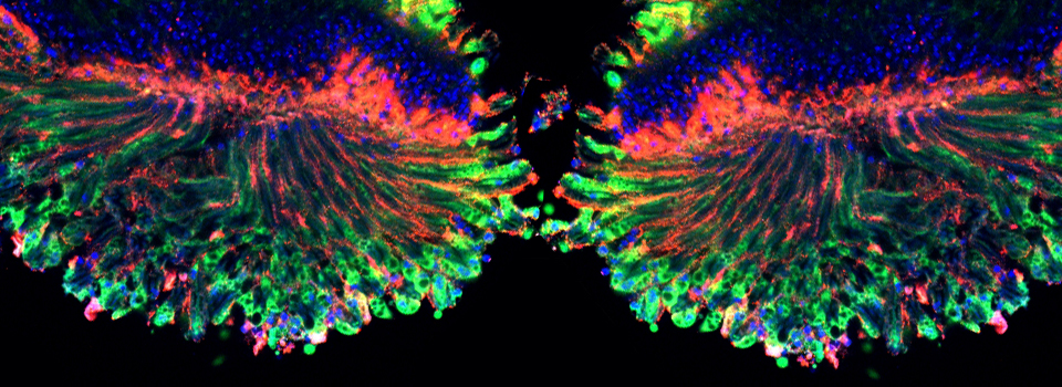
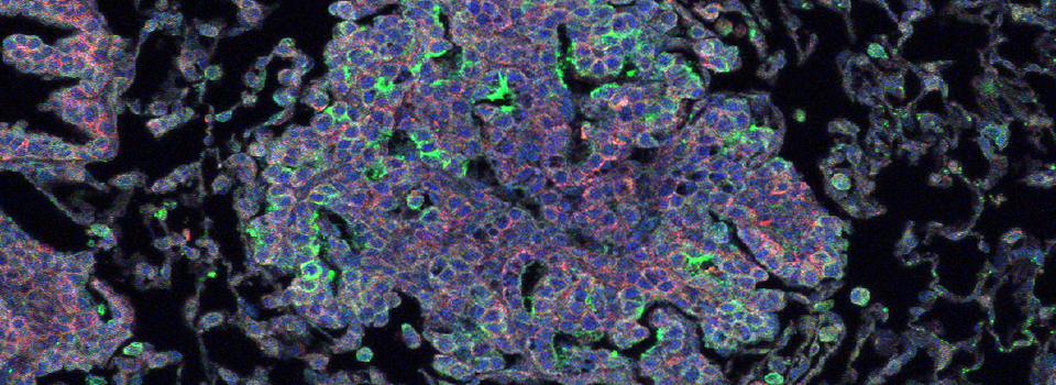
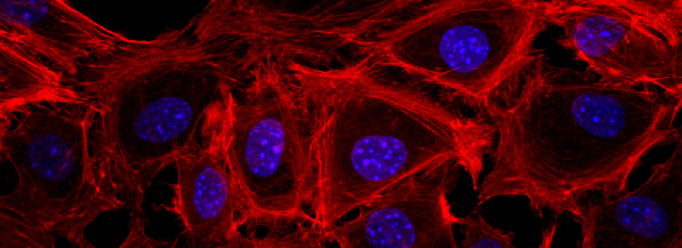
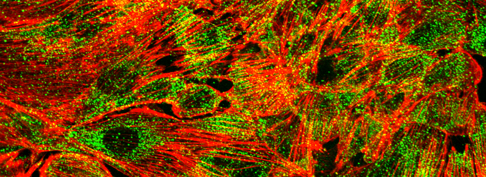
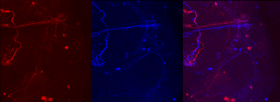
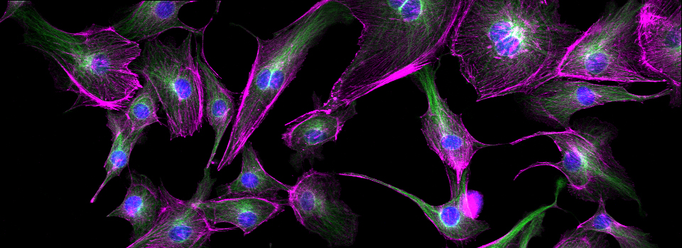
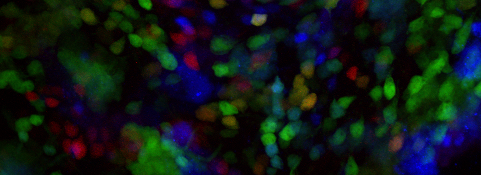
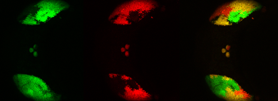
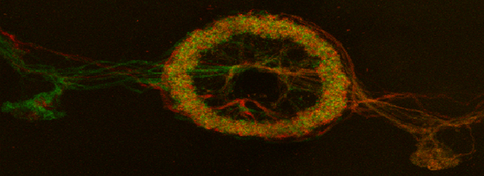
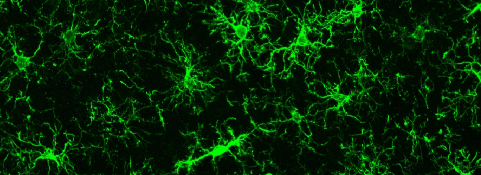
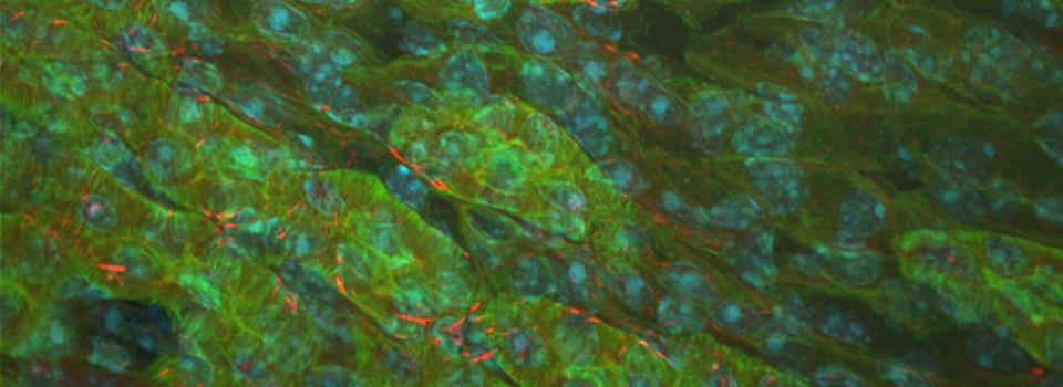
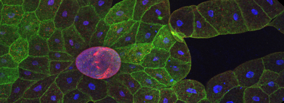
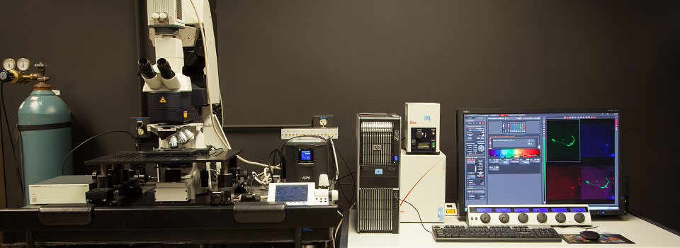
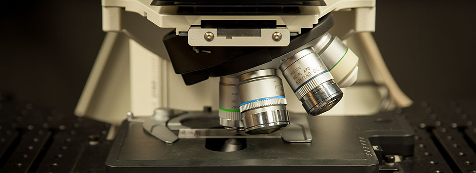
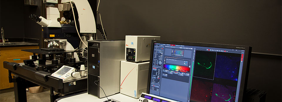
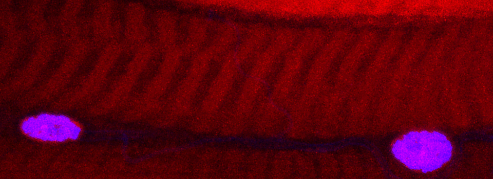
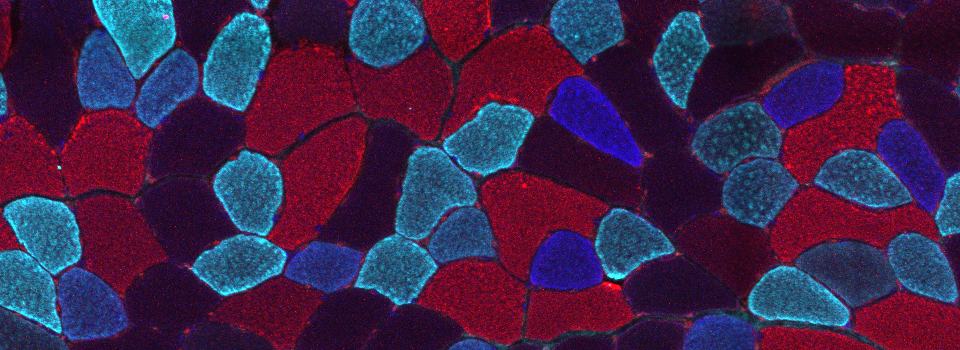
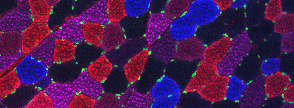
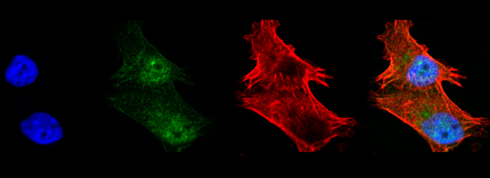

We currently have an Olympus FV3000 inverted confocal microscope (with a Tokai-Hit stage top incubator) and a Leica SP8 upright confocal microscope in our main room (SR2 328A) for taking images. We also have an Olympus inverted microscope with epi-fluorescence to allow you to check your staining quality (free of charge), a Leica MZ8 dissecting stereoscope for use in sample preparation, and a Thermo Fisher Biosafety Cabinet for putting containers with live specimens into the stage top incubator.
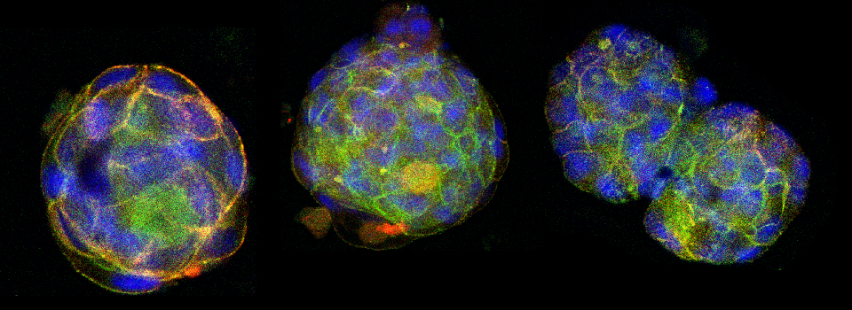
Both our confocal microscopes can simultaneously image in several different fluorescent channels as well as bright field.
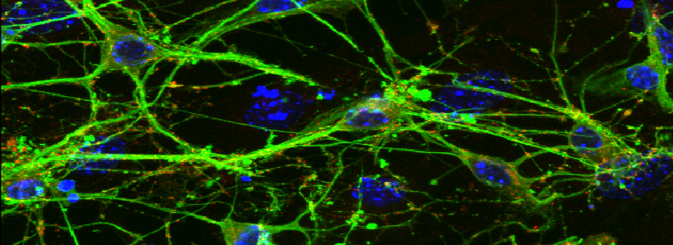
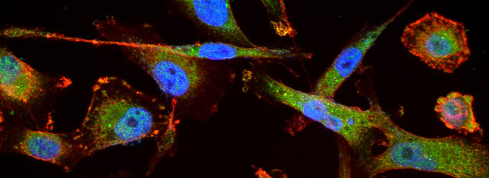
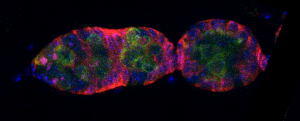
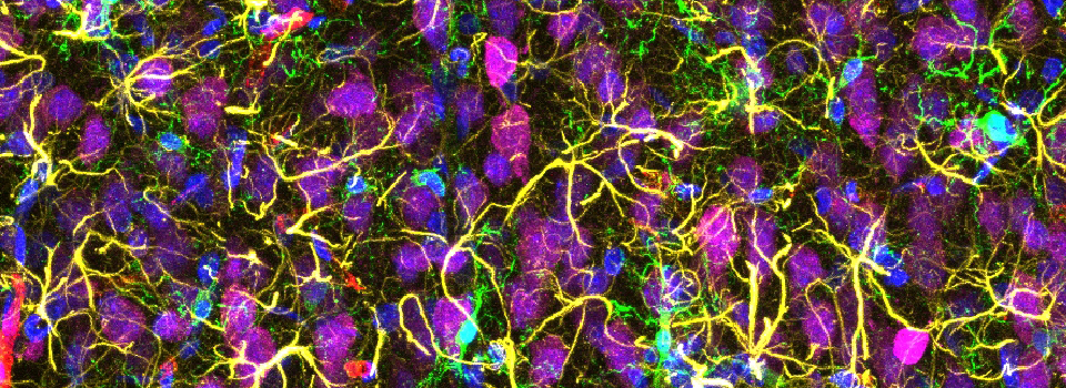
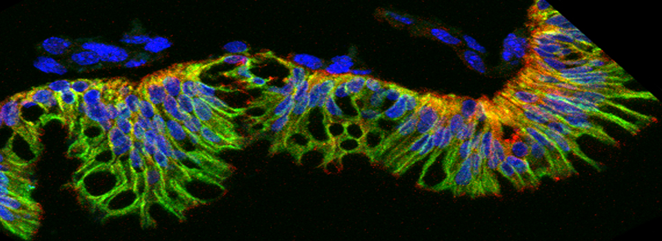
The bright multispectral capabilities of these instruments allow for imaging of multiple cell types or molecules within a sample.
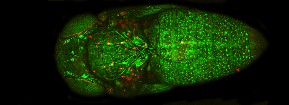
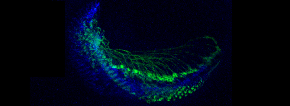
Both microscopes can perform mosaic stitching, which allows generation of complete images of samples larger than the field of view.
Both microscopes are also capable of imaging time series of living samples to capture changes in sample structure or content.
Both Microscopes are capable of beam parking, which allows one to perform experiments to measure molecular mobility such as Fluorescent Recovery after Photobleaching (FRAP). In this technique, you can photobleach areas and measure the time it takes for fluorescently labeled molecules to move back into the bleached out area.
Both Microscopes are capable of FRET measurements with a sample, which provides information on molecular proximity.