Rules & Fees
6-21-19 Changes to Data Access and Storage policy
Each PI with personnel using the BBIC microscope will have a folder assigned on the BBIC Server computer. PIs and approved personnel will have permissions to access this folder via login with their CougarNet IDs. Login via CourgarNet ID will also be used in operating the Leica SP8 and Olympus FV3000 confocal microscopes. Once a static IP address is assigned, data can be accessed/ downloaded using any computer that can connect to the UH network. Because of this upgrade, the use of external devices with the BBIC server computer is no longer allowed.
Current users (as of 6-21-19) have been entered into the new system and can access their assigned folders via login with their CougarNet IDs.
============================================================================================================
All users must be trained before they are allowed to use the confocal microscopes. This includes training on the use of the individual confocal microscopes by the Core Manager, and completion of the confocal users Laser Safety Course offered by the University of Houston (go here to check for training availability: http://www.uh.edu/ehls/training/eh23/ ). Contact the Core manager (Kathleen Gajewski, kmgajewski@uh.edu) to schedule training. You should bring some of your own specimens for your training sessions. The amount of time needed for training will vary depending on a user’s past experience with microscopy and the complexity of the planned experiments (2-4 total hours is typical). You must have the manager’s official certification as “trained” to use the microscopes without supervision. IVIS users will have additional training requirements (such as animal handling). The BBIC does not offer training in these areas, so you should contact Animal Care Operations for more information.
All users must sign up for imaging sessions via Booked. If you don’t have an account, go here:
Once you have created a user account, The Core Manager will assign you BBIC user group permissions. Besides having access to the imaging schedule, you will also be able to receive updates about the status of the equipment, any cancellations/changes in the schedule, and any other Imaging Core news of interest, which will be posted on the Dashboard. You should also e-mail your CourgarNet login to the Core Manager, so that you can gain access your Lab’s data folder on the BBIC Server.
** If you are collecting data for a project that is in collaboration with someone outside of UH, please indicate this in the notes (who you are collaborating with, and their institution(s)) when you sign up for your session. One of our funding sources requires this information. **
The first person signed up for the day is responsible for turning the microscope system on. The last person on the schedule is responsible for turning the systems off. If a system is left on overnight or the weekend, the last person on that day’s schedule will be held responsible. Repeat offenders can be denied imaging privileges.
If for some reason you cannot use your scheduled session, please go the imaging schedule and cancel it ASAP. We do understand that sometimes experiments don’t work or emergencies arise, but we do ask that you take your scheduled time off the calendar as soon as you know that you won’t be using it (a minimum of 3 hours before the scheduled start time of the next user whenever possible). If you are the last person scheduled for the day, you are responsible for notifying the core manager if you cancel to insure that the equipment is not left on overnight. If you finish your session an hour or more early, and someone is scheduled to use the microscopes after you, please ask the manager to update your end-time on the booking calendar so that the next scheduled person has the option to start earlier.
Anyone caught using the microscopes during an unreserved time can and will be kicked off the equipment. Untrained users are not allowed to use the microscopes.
During times when demand for the confocal microscopes becomes heavy, the Imaging Core reserves the right to set a maximum number of daily/weekly hours of individual use during regular business hours (M-F, 9 am- 6 pm). We understand that people can be up against hard deadlines to collect data for submitting papers/ grants/ dissertations. Please contact the Core Manager for any issues regarding user times and we will work with all parties to make scheduling arrangements that will allow everyone to meet their deadlines.
A computer workstation is available for data analysis and processing. Users should use the Booked signup sheet to guarantee use time.
External devices (hard drives and flash drives) should not be plugged into the Leica computer, as it is networked to the workstation.
Fees (starting 9/1/13):
- UH user: $25 per hour
- Outside user: $65 per hour
- Training fees: $75 per hour
- Imaging by Core: $100 per hour UH/ $140 per hour Outside
- UH user, extended time-lapse imaging into evening/weekend hours: $10 per hour during the off peak hours (6pm-9am M-F, all day Sat/Sun)
.
Logbooks will be provided for each microscope. Use a ballpoint pen, and only a ballpoint pen, to make your log entries (that means NO pencils or Sharpies!). Users should fill in ALL the information requested, especially any problems they encounter. It is especially important to fill in the start and end times. If these times differ from the start/end times on the Booked schedule, this information will allow the Core manager to make retroactive changes, if necessary. This will help ensure that you are charged only for the time you are actually using the microscopes. You are also encouraged to e-mail the Core manager about any problems or schedule changes.
Each PI will be billed once per month, and the Core manager will send an Excel spreadsheet of the lab’s usage, which is generated via Booked, to the Biology & Biochemistry accounting staff. They will send this information to NSM, which will send each PI a monthly bill. The Core maintains records of all usage and invoices, and will mail copies to any PI upon request. Non-UH users can pay via credit card using this MyNSM link.
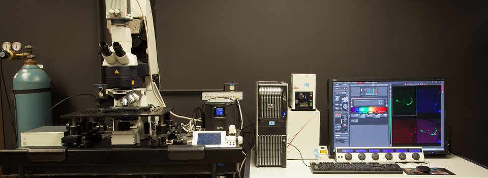
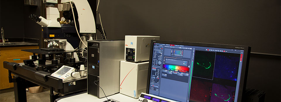
The BBIC provides a computer workstation for the processing of confocal data. It is free to use. It can handle most of the processing/analysis needs of users (exceptions are 3D manipulations for the SP8 and quantitation, deconvoluton, or image stitching for the FV3000, which can only be done on the computer that runs the Olympus microscope). Users are encouraged to do as much data processing as possible on the workstation, as use of the computers linked to the microscopes counts as billable time.
No-show rule: If you do not arrive within 30 minutes of the start of your scheduled session, or notify the Core manager in advance that you will be starting later than expected, the Core reserves the right to give your time slot to another user. You will be billed for that first half hour of wasted time.
Service visits: Our microscopes are very complex pieces of equipment, and complex equipment requires regular maintenance. We will do everything in our power to schedule service visits so that they do not take away from anyone’s reserved use times, but sometimes we are limited by the individual engineers’ work schedules. In those circumstances we may have to pre-empt someone’s scheduled time. We ask our users to be patient when we must take away scheduled imaging time for service visits, as it is in everyone’s best interests to have our instruments in proper working order. You will be informed via e-mail as soon as we confirm the visit with the engineer, and you will not be billed for any time that must used for service/repairs.
After Hours Access: All UH members of the Biology Imaging Core group may request card access to SR2 328A. You should e-mail the Core manager Dr. Kathleen Gajewski ( kmgajews@Central.UH.EDU ), provide your 7 digit PeopleSoft ID#, and request to be included on the card reader list. People who are still in the process of being trained cannot use the microscopes after hours.
The Core door is locked for a reason, and THIS DOOR SHOULD NEVER BE PROPPED OPEN WITHOUT PERMISSION FROM THE CORE MANAGER OR CORE PI. NO EXCEPTIONS!!
Use and Care of Equipment
If you are using the mercury lamps (which you will if you want epifluoresence), once they are switched on they need to remain on for a MINIMUM of 30 minutes. Once they are switched off they need to remain off for a MINIMUM of 30 minutes. This will help to extend the operating life of the bulb. If someone is scheduled to use the microscope within 3 hours of the end of your session, leave the mercury lamp ON. The mercury lamps are labeled box #1 for both microscopes.
The time booked on the microscopes should be used for imaging/data collection. We have a separate workstation for data analysis and processing.
Do not leave your images on the Olympus FV3000 computer hard drive for long term storage. Files are automatically saved to the D drive, but you should copy them to your lab’s folder in the BBIC server after your session, and delete them from the FV3000 computer. Image files can be very large and will quickly bog down the computers. Data left on the hard drives is subject to erasure. All data collected on the Leica should be saved directly to the networked Computer Workstation. Leaving a small file or two in a clearly labeled folder on the confocal computer hard drives as references for future imaging is permissible with approval of the core manager.
Be considerate of the equipment. Clean the oil/other fluids off the objectives after you are finished (even if someone is scheduled after you). This is especially important for the FV3000, as it is an inverted microscope, and it is all too easy for excess oil to start running down the sides of the objectives. Should you accidentally get oil on a non-oil objective, wipe off what you can with lens paper and tell the core manager ASAP so that the lens can be cleaned promptly. If you are the last user of the day, follow the shutdown protocols and leave the fans (green “Laser Power” button on the SP8) running for at least 5 minutes (this will help to extend the operating life of our argon lasers). Do not leave the equipment on overnight. Please cover the microscopes if you are the last user of the day. Abuse/neglect of the microscopes can lead to loss of imaging privileges.
Each microscope has its own supply of immersion oil (provided by the manufacturers). You should use this oil, AND ONLY THIS OIL, for your imaging. Different oils have different indices of refraction, and mixing such can result in a blurry mess and lost imaging time while objectives are cleaned. If you already have oil on a slide, and you’re not sure it will be compatible, clean it off with 95% ethanol before you put it on one of the microscopes. Lens paper is provided for cleaning the objectives; USE THIS PAPER ONLY. Also note that the FV3000 has both Silicon Oil objectives (green oil bottle), as well as standard oil objectives (blue oil bottle)- double check that you have the right bottle before oiling an objective!
Users should do as much sample prep as possible within their own labs. We do have bench space and a dissecting microscope for work that needs to be done near the microscopes. Users should bring their own equipment/ reagents, and clean the area after they have finished. Slides brought in for imaging MUST have their coverslips completely sealed with nail polish so that no mounting media is leaking out from under the coverslip.
Users should log their imaging times and any issues with the equipment in the logbooks next to each microscope. This will insure accurate billing for actual time used, and documentation of any problems.
Users having problems with the equipment should also send the Core manager an e-mail with a detailed description of the equipment issues.
Unconventional experimental setups: If your experiment requires major adjustments to the stage and/or setting up additional equipment, you are responsible for returning the microscope and its surroundings to their original state by the end of your scheduled imaging time (so allow for this break down time if someone is scheduled after you). If you need to take objectives off a microscope for any length of time for any reason, you must put them in their cases (which are in a drawer in the back of the room marked “Leica hardware”) and set them on the back counter out of harm’s way. Please insure that they are returned to their original positions (see #12) on the microscope.
Leica stages: We have two types of stages available for use with the SP8, a motorized stage (which is required for mosaic imaging), and a large translational stage (which can be used for electrophysiology set-ups, and for live-imaging larger specimens, such as rodents). For many applications, either stage will work just fine. If you require a specific stage, and it is not currently mounted on the Leica microscope, you must contact the Core manger at least several days in advance and request a stage swap. The swap can take 30-60 minutes, so we must reserve the necessary time. We want to accommodate everyone’s imaging needs, and if it becomes necessary we may schedule certain weeks for one stage or the other.
The objectives on the Leica SP8 must ALWAYS be changed BY HAND.
When the translational stage is in use it can be raised/lowered by hand, but there is no way to communicate that change to the software option for controlling the objectives. This could lead to the dreaded objective-slide collision, or the even worse objective-stage collision. When the scanning stage is on, the stage insert has raised buttons that can sometimes be in the path of the objectives. The software doesn’t know this either, and can’t compensate; YOU, the human user, can. Therefore change them by hand, NO exceptions.
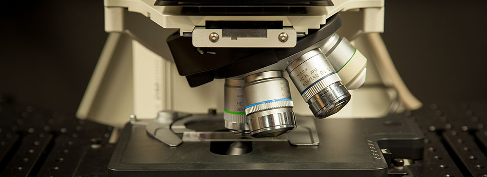
Leica objectives: Our SP8 can hold 6 objectives at a time. However we currently have 8 available, and are always looking to add more types of objectives to expand our users’ options for collecting data. Each turret position is programmed for a specific objective. It is VERY IMPORTANT to put the objectives in the right spots, as optimal confocal performance depends on precise engineering on the light paths, which in turn depends on the system recognizing the objective in use. This is the default objective assignment list:
Position 1: 20x- N/A 0.70, dry
Position 2: 10x- N/A 0.30, dry
Position 3: 40x- N/A 1.30, oil
Position 4: 63x- N/A 1.40, oil
Position 5: 25x- N/A 0.95, water
Position 6: 100x- N/A 1.40, oil
The number to the left of the objective denotes the turret position:
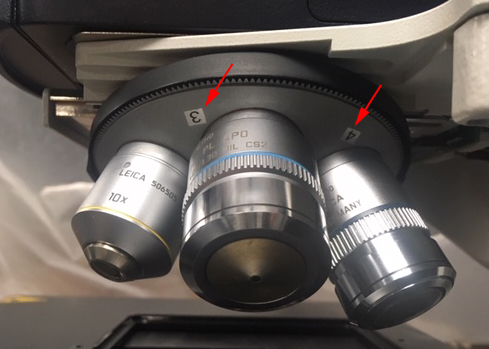
If you wish to use an objective not currently mounted on the SP8 or FV3000, see the manger first. The software will need to be reset to recognize the new objective, and only the manger or Core PI are allowed to make such adjustments.
Laser Safety Protocols
Laser Safety Standard Operating Procedures:
- Follow approved procedures for turning on and turning off lasers.
- Do not look through microscope eye pieces while laser is scanning sample.
- Keep your hands and face away from the sample/ stage area while the lasers are active.
- Never place reflective materials such as mirrors, chrome or steel instruments near sample while scanning.
- Do not scan any samples that contain dyes capable of generating toxic airborne contaminants when irradiated by lasers. If you do not know, do not use!
- Never handle the laser housing.
- Alignment of the lasers will only be performed by the microscope manufacturer’s representatives.
- If your eyes are exposed to laser irradiation, seek medical attention immediately. Then contact the Core Manager, Core PI, and UH Radiation Safety Officer as soon as possible.
- All users are required to take the UH Environmental Health & Safety course on laser safety (EH23) and take a yearly refresher course (EH23W). These courses are offered in person several times a year, and notices are posted on the Booked dashboard. The refresher course can also be completed online.
Your Responsibilities as a User:
At the risk of being redundant, here is what we expect from our users:
Clean up after yourself- leave the microscopes/ benches the way you found them (and if you found them dirty- notify us!).
Stay informed- Everyone who uses the Core is required to have a Booked account, and any Core updates are posted to the dashboard by the Core manager. These updates are IMPORTANT and there is no excuse for failing to read them. You are responsible for keeping up to date on Core news- Read the Dashboard posts.
Report any problems- If you are having troubles with your imaging, go find the manager! Often the problem is something simple that can be figured out and fixed easily. If not, we need to know about it so that we can bring in someone who can fix it. Any issues should also be recorded in the logbooks.
Play nice and share- If there are scheduling problems, talk to the manager first about them! We expect to have many users, and no one has any more right to the equipment than anyone else. We will give priority to users with deadlines, but if you need a large amount of time on these instruments, you will be expected to also make use of evening and weekend time slots, so that others may have access during regular business hours. Remember that most experiments can be done equally well with either confocal microscope.
Help the Core run smoothly- Please give us all the necessary information about your sessions. Fill in the logbooks and the online scheduling windows. Notify us promptly if you must change your scheduled use times. Check the schedule often on days you use the Core, in particular AFTER your session. Remember, it can be changed in a moment’s notice.
