Which objective is best for me to use?
Our confocal microscopes are equipped with several different types of objectives. Each one will have its own strengths and weakness, as there is no universal objective that is optimal for all types of experiments. The choice of the one that works best for your planned experiment will be greatly influenced by the type of specimen and how it is mounted.
Three of the most important objective properties to consider are 1) the Numerical Aperture (N/A), 2) the immersion media, and 3) the Free Working distance.
The N/A number is a measure of an objective’s resolving power, i.e. the shortest distance between two points on a specimen that can still be distinguished as separate objects. The numbers on our objectives range from 0.08 to 1.4; the higher the number, the greater the resolving power. N/A is determined by the objective’s capacity to collect the available light- the more light collected, the greater the resolution. Diffraction of the light as it passes through different media (glass, air, water, tissues, etc.) reduces the resolution. This loss of light can be reduced by using objectives with different immersion media.
As the N/A number of an objective increases, other parameters such as working distance and the area of the field of view tend to decrease, which could be relevant in conducting your experiment.
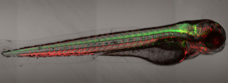
The immersion media refers to the substance that is touching the objective lens when it is in use. We have 4 types: air, water, standard Immersion Oil, and Silicon Oil. Air objectives have the lowest N/A ratings, as there is much diffraction of light that passes from the objective lens, through air, then through the coverslip. These are not the preferred objectives to do detailed imaging of sub-cellular structures. But they are useful for images of larger specimens such as zebrafish embryos, and they are also needed for applications where it’s not possible to use another immersion medium, such as trying to take pictures of all the wells in a 96 well plate on MATL mode (where the stage is programmed to move to each well). Water objectives are designed to dip directly into an aqueous medium. They have higher N/A ratings than air objectives, but are not as powerful as oil objectives. Immersion oil objectives work with oil that has a refractive index closest to that of glass (>1.5), and have the greatest resolving power. The Si oil has a lower refractive index, closer to that of living tissue. This oil is also more stable than standard immersion oil, which makes the SI oil objectives a good option for extended time lapse imaging of live specimens.
The working distance (the distance between the front edge of the objective lens and the specimen surface with the surface of the cover glass) can be looked at as how far an objective can “see”. The higher the N/A #, the more “near sighted” an objective tends to be, i.e., the smaller the working distance. If the desired focal plane lies beyond the objective’s listed working distance, no amount of turning on the focus knobs will bring that image into focus. If you are using the Leica SP8 (the upright confocal microscope), you risk ramming the objective through your slide (a bad outcome to be avoided) or otherwise dislodging/damaging your specimen. If you are using conventional mounting devices (slides, dishes, well plates) for your specimens, you will usually not have issues with being in the range of the working distance. But if you are using something more unusual (such as microfluidic chips), you will need to consider the working distance and how your specimen fits between the objectives and the stage.
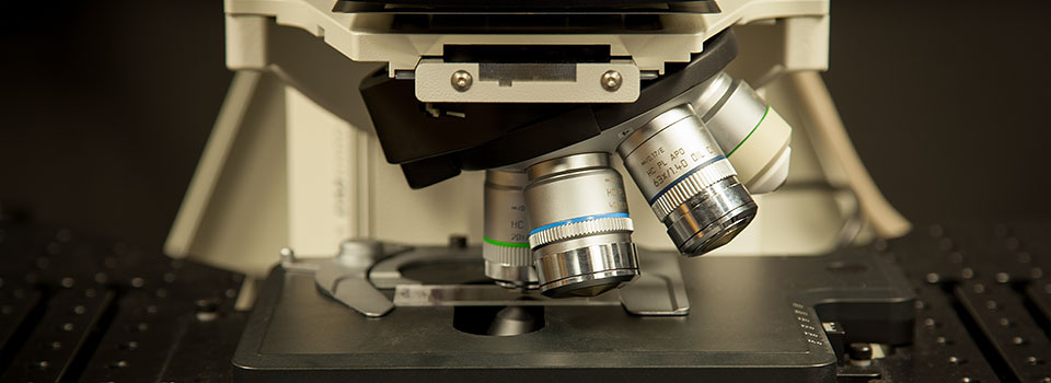
Another useful feature on some objectives is a correction collar. This is a dial on the tip of the objective that allows the user to realign the focal points of the light rays. This is especially needed with coverslips that are thicker or thinner than the #1.5 type ( ideally 170 μm, but they can range from 160-190 μm) that confocal systems are engineered to use. On the example below, the correction collar has 0.17 (for a #1.5 coverslip) lined up with the tick mark (yellow arrow). If you had a slide with a #1 cover slip (150 μm), you can turn the dial to put the tick mark between 0.13 and 0.17 to see if you can get a focus on your specimen.

3 of our FV3000 objectives have this feature. The microscope also has a correction collar adjuster, so that you can make adjustments to the dial without having to remove your specimen.

Most of the objectives in our collection can be used for multiple types of experiments. Some are specialized for specific types of tasks.
Air objectives
FV3000 2X PLAN APO: This objective can scan an entire well of a 96 well plate, so it is ideal for a quick scan. It is also useful for making a reference map of a very large specimen.
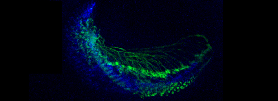
FV3000 10X UPLSAPO10X2: This objective is mostly used for getting an initial focus on a specimen before switching to a higher mag objective, and for making reference maps. It also produces very good pictures of later stage zebra fish embryos, and is handy for imaging them during Multi Area Time Lapse (MATL) experiments.

FV3000 20X UCPLFLN20X: This is the objective for MATL scans of well plates that would go beyond the range of an oil drop. It also has the special feature of being compatible with plastic-bottomed plates and dishes.
SP8 HC PL Fluotar 10x: This objective is mostly used for getting an initial focus.
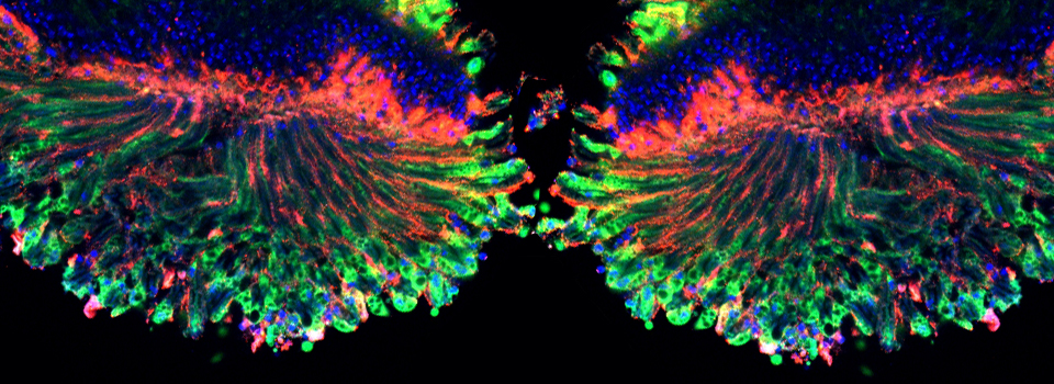
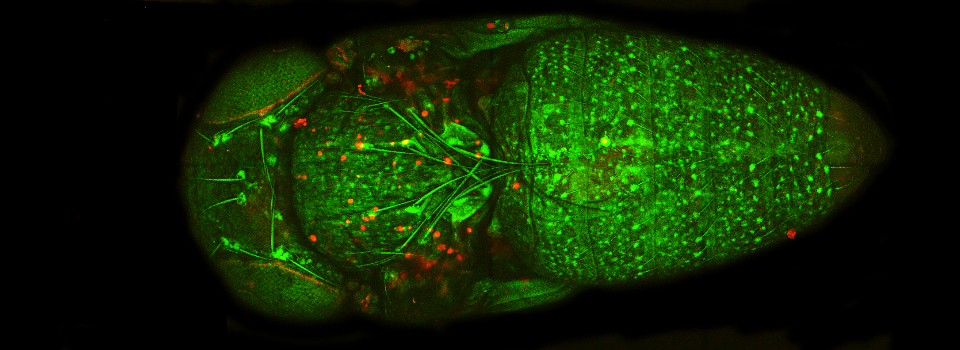
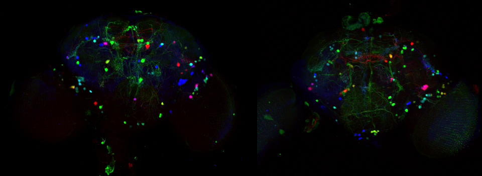
SP8 HC PL APO 20x: This objective has a very high N/A (0.75) for an air objective, and is ideal for imaging a whole specimen the size of a Drosophila brain.
Water objectives
SP8 HCX IRAPO25x, SP8 HCX APO L U-V-I 40x, SP8 HCX APO 63x: These objectives are ceramic instead of metal, so they are ideal for electrophysiology experiments.
Silicone Oil Objectives
FV3000 30X UPLSAPO30XS: This objective has an very long working distance (0.8 mm!) for its N/A number (1.05) and is ideal for imaging thicker specimens such as zebra fish embryos and brain explants.
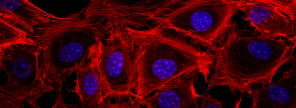
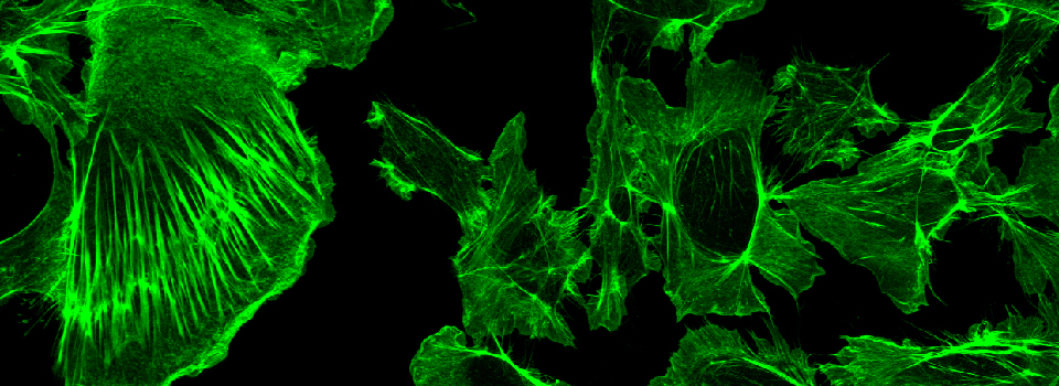
FV3000 40X UPLSAPO40XS: This objective is the best choice for longer time lapse imaging of live cells in culture.
Immersion Oil Objectives
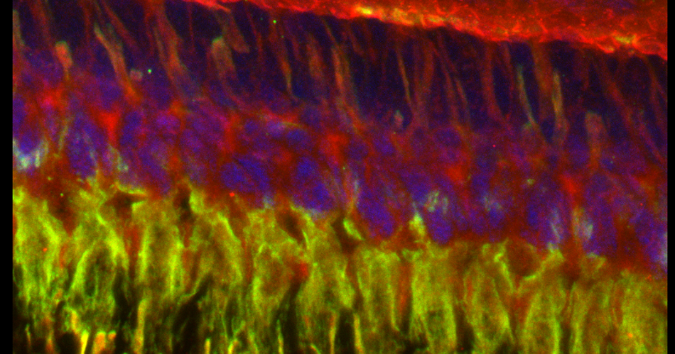
These objectives will have the highest N/A scores (starting at 1.3), and are the best choices for imaging down to the level of large sub-cellular structures.
FV3000 60X PLAPON60XOSC2
This objective is said to have the absolute best correction for chromatic aberration, and should be your first choice for doing co-localization analysis.
SP8 HC PL APO CS2 40x, SP8 HC PL APO CS2 63x, SP8 HC PL APO CS2 100x
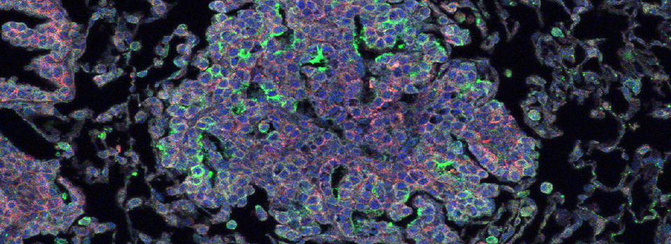
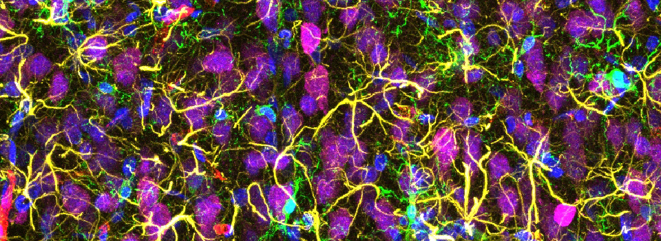
The 40x is a popular choice for big (>20 stacks) tile scans of larger specimens such as rat brain sections, as it offers a good balance between resolving power and field of view size (which contributes to scan times). The 63x and 100x offer the higher N/A, at 1.4.
MANUFACTURER’S SPECS ON LEICA SP8 OBJECTIVES

MANUFACTURER’S SPECS ON OLYMPUS FV3000 OBJECTIVES
AIR OBJECTIVES

SI-OIL OBJECTIVES

OIL OBJECTIVE
