Data from the Demo of the Nikon BioStation CT
This instrument is designed for doing long-term (as in days, weeks, or even months!) time-lapse imaging of live cells in culture that would be expensive and impractical to do with a standard confocal system. This type of kinetic imager would complement our confocal microscopes, as it can do longer term, as well as high volume time courses. Its large capacity makes it ideal for conducting large scale drug screening and also allows multiple experiments to run simultaneously. We have hosted 2 demos of this instrument, and have plenty of data to show any interested parties. This acquisition is currently tabled, but should there ever be sufficient interest in the UH research community, we are ready to seek a funding source to obtain it.
For more information about the BioStation, go here:
Here is a movie of the BioStation robot arm in action, moving a plate to the carrier:
Here are some of the images collected by our researchers during the demo:
1) Natural Killer Cells vs. K526 cells
(Lyle Babcock and Austin Bigley, Mark Clarke’s Lab, Dept. of Health and Human Performance, University of Houston)
These images are a crop from a centered tile scan on a 6 well plate, taken every 10 minutes (20x objective). Here you can clearly see several K526 cells swell up and burst as a result of attack by the NK cells.
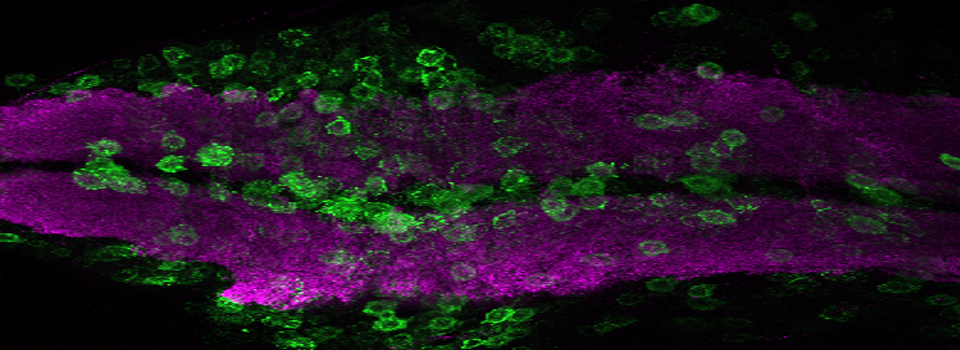
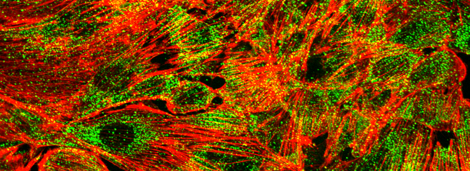
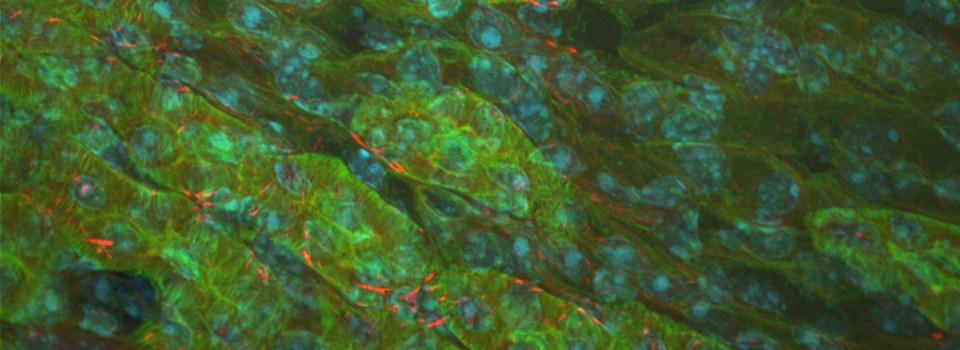
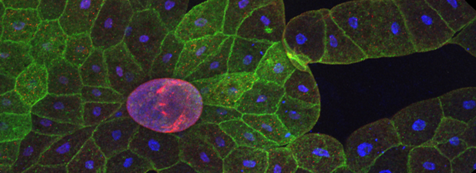
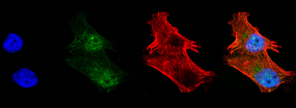
2) PC3 Prostate cancer cells
(Anders Strom, CNRCS, Jan-Åke Gustafsson’s Lab, University of Houston)
This is a portion from a tile scan (10x objective) of PC3 cells transfected with GFP-HIF-1alpha and mCherry-C1, imaged every 4 hours over 2 days, using phase contrast and 2 fluorescent channels.
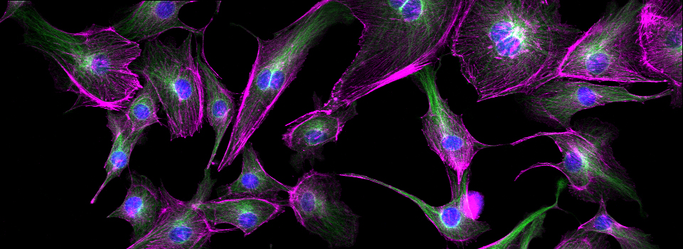
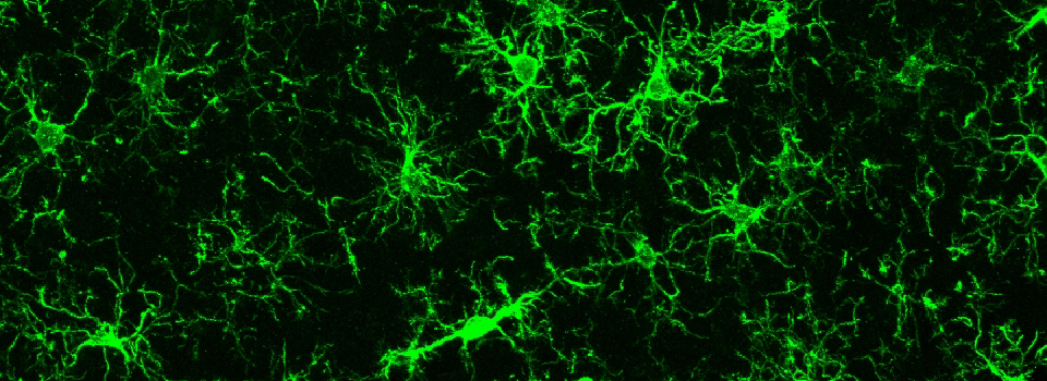
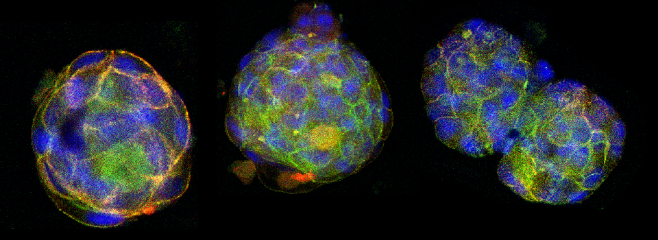
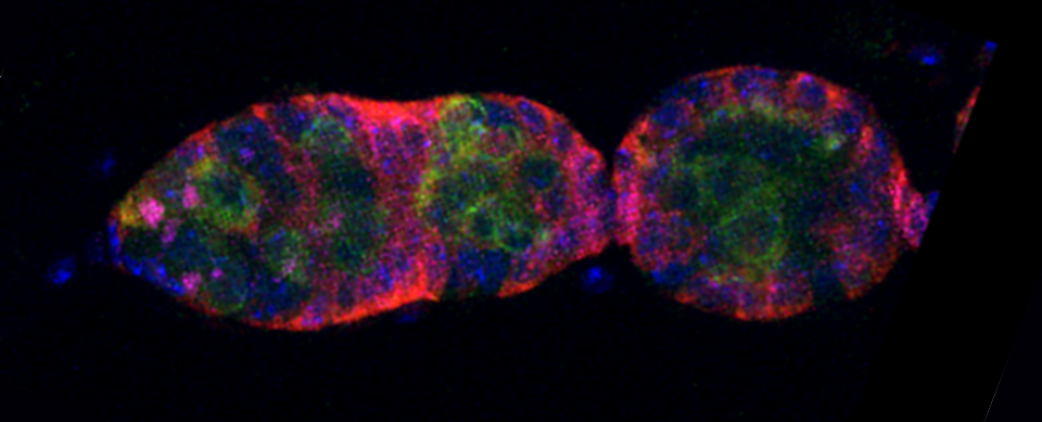
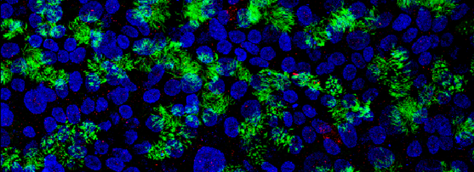
3) Mammospheres growing and merging in culture
(Santosh Kumar, Tasneem Bawa’s Lab, CNRCS, University of Houston)
This is a portion from a tile scan (10x objective) imaged every 3 hours.
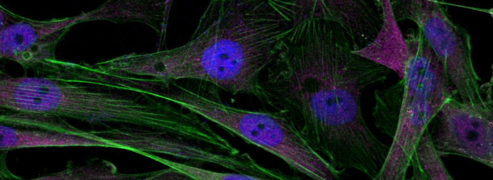
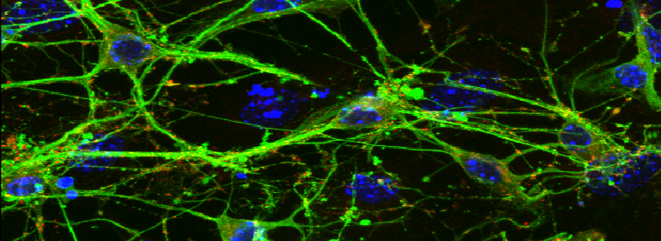
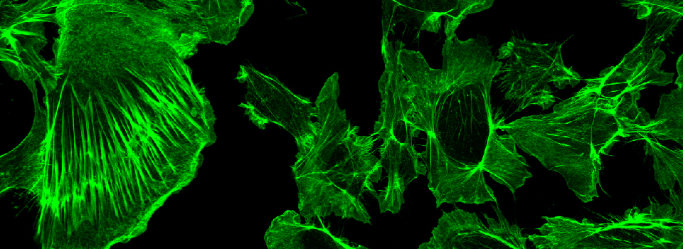
4) Scratch migration assay with mda ampk cells
(Efrosini Cuko, Dan Frigo’s Lab, CNRCS, University of Houston)
This is a scan of a pre-selected point imaged every 30 minutes (10x objective).
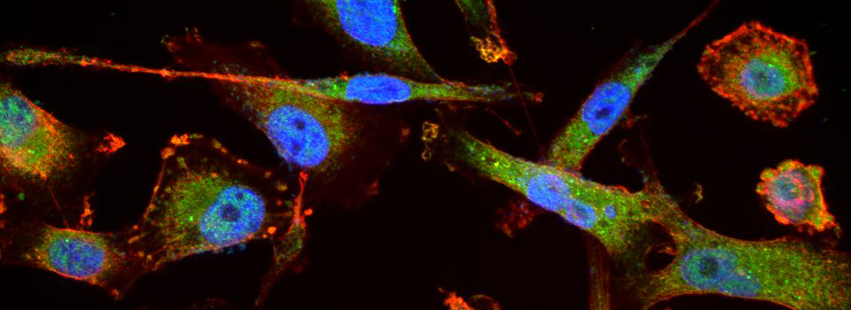
5) Scratch migration assay with sum159 cells
(Efrosini Cuko, Dan Frigo’s Lab, CNRCS, University of Houston)
6) An Apoptosis Assay
(Joseph Manarang, Gomika Udugamasooriya’s Lab, Department of Pharmacological & Pharmaceutical Sciences, University of Houston)
Going off the beaten path- unconventional applications for the BioStation.
The BioStation was designed with cell cultures in mind, but we also tested other experimental systems, with an eye towards expanding its potential users. Our results show the possibilities of imaging larger specimens such as Drosophila and zebrafish.
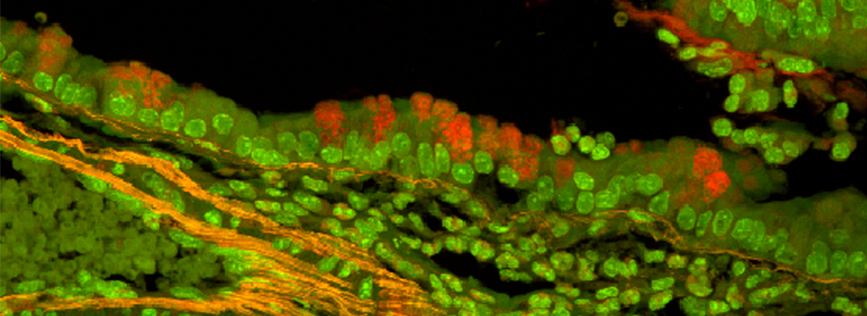
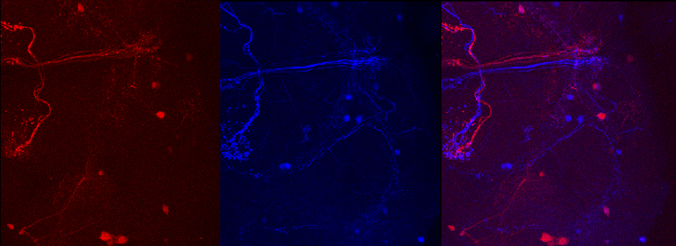
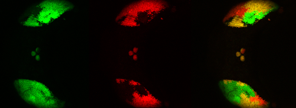
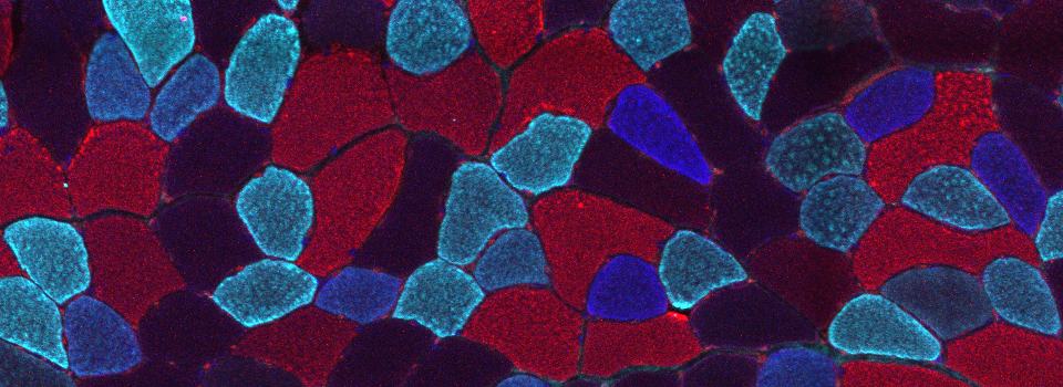
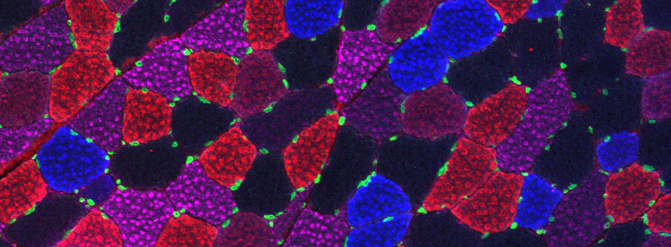
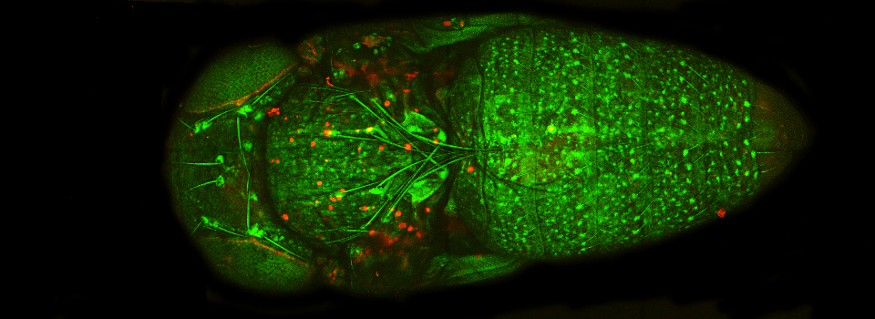
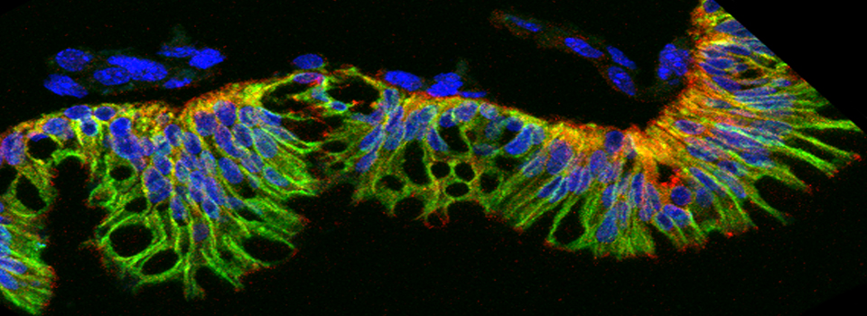
1) Early metamorphosis of a Drosophila pupa
(Kathleen Gajewski, BBIC, University of Houston)
This animal is expressing GFP and/or RFP from Brainbow, in the eye imaginal discs (gmr-GAL4 driver). This fluorescence allows tracking the migration and growth of these tissues as they form the compound eyes and the ocelli. Preselected point scans were taken every 10 minutes, using phase contrast and 2 fluorescent channels (4x objective).
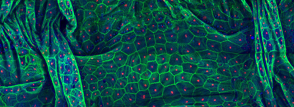
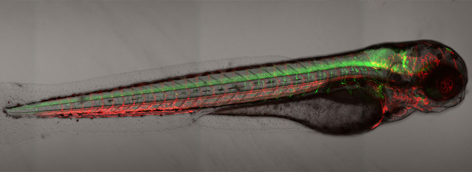
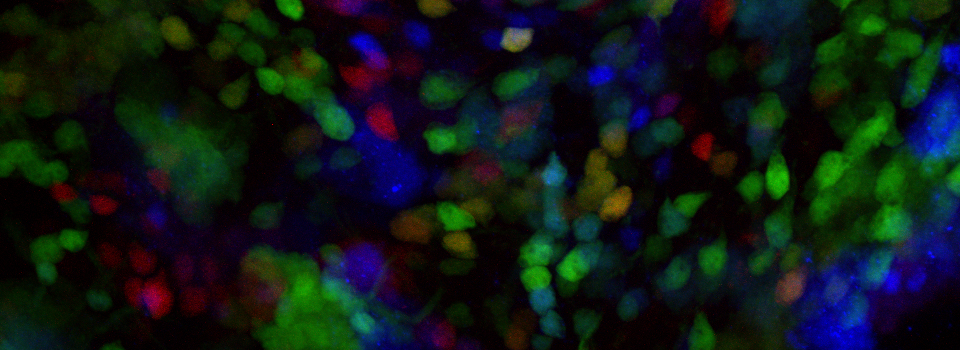
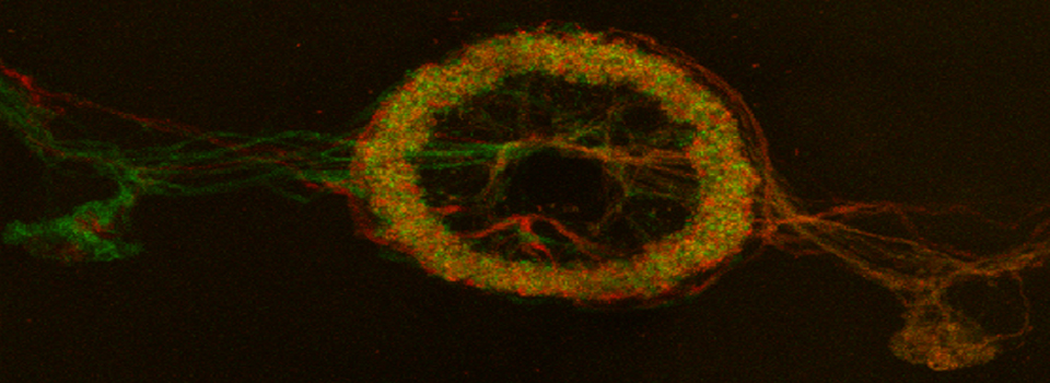
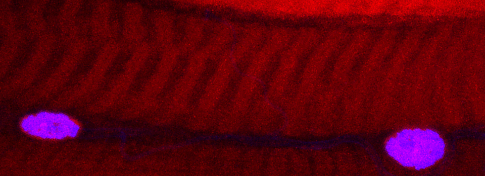
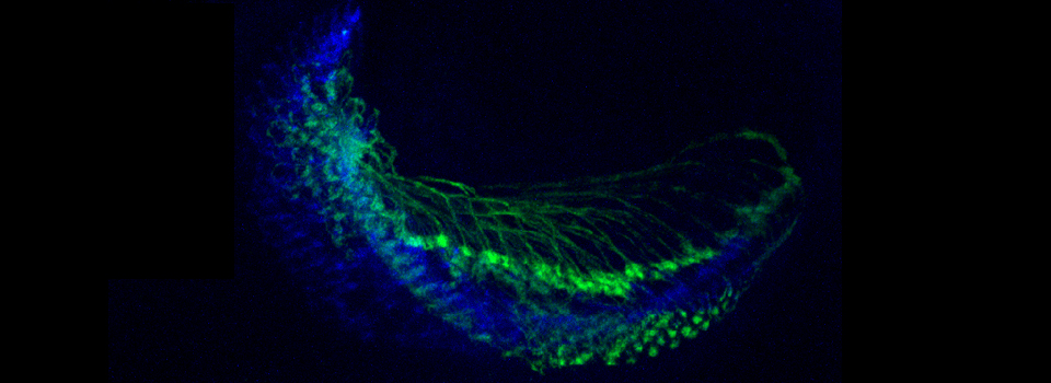
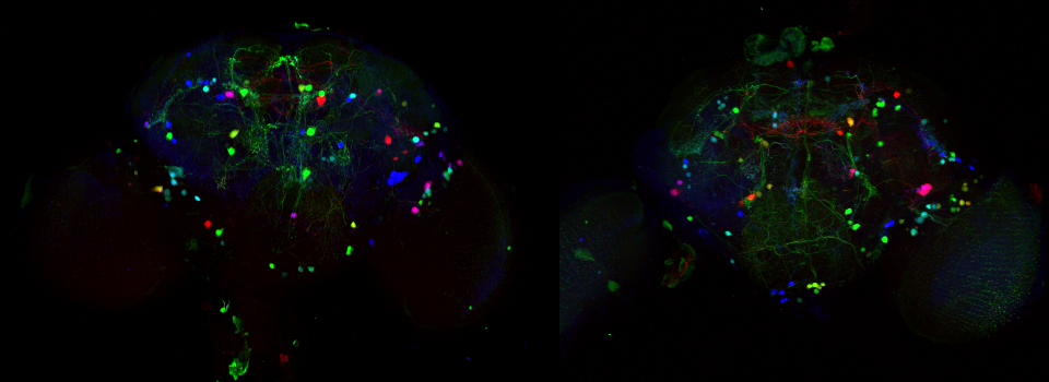
2) Zebrafish embryo
(Maria Bondesson, University of Houston)

This is a single whole well scan (4x objective) of a Zebrafish embryo expressing SAGFF(<F)53A, a random transgenic insertion that drives GFP in the developing skeleton.
If you did not have a chance to try the BioStation, but you have current or future projects that would benefit from this technology, please contact us. The more interest we can demonstrate from the UH research community, the better our chances of getting funding.